In this article, Steven Fleming PhD, our Director of Embryology, discusses the nature of GM-CSF, the rationale for using it during extended culture in vitro, and its possible clinical benefits for frozen-thawed embryo transfer.
What is the role of GM-CSF in reproduction?

Figure 1: Embryo-maternal intercellular communication during embryogenesis and implantation
GM-CSF is one of a family of pleiotropic cytokines believed to be involved in the regulation of diverse reproductive functions including gamete maturation, ovulation, embryogenesis, implantation, pregnancy, and parturition.1
Cytokines are potent cell-secreted intercellular signalling proteins that may have an autocrine, juxtacrine, paracrine, or endocrine effect at nanomolar to picomolar concentrations, binding to specific cell membrane receptors to activate intracellular signalling pathways.
Indeed, there is constant embryo-maternal communication that maintains embryo viability and regulates implantation (Figure 1). Interestingly, GM-CSF can upregulate its own expression within the endometrium, suggesting that it also has an autocrine effect within the uterus.2
The rationale for adding GM-CSF to culture media
To justify the addition of GM-CSF to culture media, it is mandatory to verify its secretion into the female reproductive tract. Moreover, it is necessary to demonstrate the presence of GM- CSF receptors on embryos and, likewise, the dependence of embryo survival and viability upon GM-CSF.
In this respect, GM-CSF has been shown to be expressed, synthesised, and secreted from the human oviductal and endometrial epithelium.2-4 Furthermore, levels of GM-CSF reach a peak within the reproductive tract during the secretory phase of the menstrual cycle4,5, which coincides with the time of conception and implantation.
The GM-CSF receptor is a dimer comprised of a cytokine-specific alpha subunit (GM-Rα) and a signal transducing beta subunit (βc), βc forming a high-affinity complex with ligand-coupled GM-Rα.6 However, GM-Rα can also act alone, for example, to mediate the effect of GM-CSF on glucose transport via a kinase-independent pathway.7 Indeed, GM-CSF has been shown to promote glucose transport in the embryo, which expresses the GM-Rα receptor throughout preimplantation development.8,9
A beneficial role of GM-CSF during embryogenesis was first demonstrated over 30 years ago in the mouse.10
In a knock-out mouse model, GM-CSF was shown to be obligatory for normal embryo development, implantation, and fetal viability.11
Furthermore, the addition of recombinant human GM-CSF (rhGM-CSF) to culture media has been shown to enhance human blastocyst development and viability.9,12
The central concept of GM-CSF as a survival factor
During preimplantation embryogenesis, the embryo responds to a variety of growth factors and cytokines, some of which act as mitogens, stimulating cell proliferation, others which act as survival factors, preventing programmed cell death, also known as apoptosis.13
In this respect, particularly during assisted reproduction in vitro, embryos may be exposed to a multitude of stresses which can result in developmental arrest and even cell death.
However, survival factors manage to mitigate the effect of cellular stressors via the phosphoinositide 3-kinase (PI3 Kinase) pathway and thereby can increase embryo viability (Figure 2).

Figure 2: Mitogens, survival factors and cell stress13
Indeed, using the mouse model, the potential benefit of co-culture with Vero cells and culture media containing a wide variety of growth factors and cytokines to embryo development following cryopreservation at the 1-cell stage, revealed GM-CSF to be the only one capable of enhancing cell survival and preventing apoptosis (Table 1) though, notably, it did not influence overall cell number within the developing blastocyst.14
Table 1: The impact of various culture conditions on embryogenesis and apoptosis14
| Treatment | Blastocyst rate (%) | Hatching rate (%) | Cell count (Mean) | Apoptotic index (Mean) |
|---|---|---|---|---|
| Control | 89 | 59 | 100±44 | 0.50±0.68 |
| Vero cells | 87 | 78 | 124±42a | 1.70±1.29b |
| GM-CSF | 80 | 69 | 100±46 | 0.00±00c |
| LIF | 87 | 68 | 99±41 | 0.67±0.81 |
| TGF-α | 87 | 84 | 108±40 | 0.69±0.87 |
| IGF-I | 87 | 76 | 105±39 | 0.23±0.43 |
| IGF-II | 93 | 72 | 102±39 | 0.28±0.50 |
| FGF-4 | 85 | 65 | 106±40 | 0.49±0.72 |
| TNF-α | 79 | 67 | 119±38 | 0.18±0.30 |
| TGF-β | 79 | 65 | 100±41 | 0.69±0.78 |
| IL-6 | 80 | 74 | 113±39 | 0.68±0.88 |
| PDGF | 88 | 82 | 103±43 | 0.45±0.85 |
| EGF | 91 | 56 | 84±47 | 1.23±1.49 |
a,b,cSignificantly different from control (p < 0.05).
cNo apoptotic cells observed with GM-CSF-treated embryos.
Interestingly, a retrospective, case-controlled study of 39 couples whose embryos had repeatedly suffered severe cleavage arrest during two previous IVF cycles, subsequently experienced perfectly normal blastocyst development when their embryos were cultured in medium containing GM-CSF, resulting in implantation, clinical pregnancy, miscarriage, and live birth rates entirely consistent with a control group of 80 first cycle tubal-factor patients (Table 2).15
Table 2: Cycle characteristics and pregnancy outcomes in GM-CSF and Control groups15
| GM-CSF group (n=39) | Control group (n=80) | p | |
|---|---|---|---|
| Antagonist protocol (n, %) | 23/39 (59) | 55/80 (69) | 0.309 |
| Total r-FSH dose (IU) | 2834 (1875-3375) | 2576 (1600-3206) | 0.290 |
| Cycle cancelation rate (n, %) | 5/39 (12.8) | 9/80 (11.2) | 0.802 |
| Number of retrieved oocytes | 6.3±3.0 | 7.0±3.0 | 0.292 |
| Number of M-II oocytes | 4.9±2.6 | 5.6±3.5 | 0.263 |
| Number of 2PN | 3.7±2.0 | 3.9±2.5 | 0.576 |
| Number of embryos transferred | 1.3±0.5 | 1.3±0.5 | 0.707 |
| Embryo transfer day (n, %) | – | – | 0.416 |
| Day 3 | 11/34 (32.4) | 29/71 (40.8) | – |
| Day 5 | 23/34 (67.6) | 42/71 (59.2) | – |
| Implantation rate, (%) | 32.3±44.2 | 33.1±46.2 | 0.937 |
| Clinical pregnancy rate, (n, %) | 13/39 (33.3) | 26/80 (32.5) | 0.770 |
| Miscarriage rate, (n, %) | 3/13 (23.1) | 5/26 (19.2) | 0.997 |
| Live birth rate, (%) | 10/39 (25.6) | 21/80 (26.2) | 0.943 |
Data are expressed as means ± SD, p<0.05, median (min-max) and percentages.
GM-CSF: Granulocyte-macrophage colony-stimulating factor, r-FSH: Recombinant follicle-stimulating
hormone, M-II: Metaphase II, SD: Standard deviation
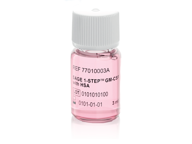
The clinical benefit of SAGE 1-Step™ GM-CSF medium
SAGE 1-Step GM-CSF medium may be used for various purposes, including extended embryo culture, recovery culture following blastocyst vitrification and warming, and as an embryo transfer medium.
A longitudinal, retrospective, propensity matched, cohort study analysed clinical outcomes following extended culture in Naka ONESTEP medium, resulting in vitrification of suitable blastocysts. Recovery culture in SAGE 1-Step GM-CSF medium or control Naka ONESTEP medium was performed for 3-5 hours post-warming prior to frozen blastocyst transfer.16 The rates of positive human chorionic gonadotrophin (hCG), biochemical pregnancy, clinical pregnancy, ongoing pregnancy, and live birth were all significantly higher in the SAGE 1-Step GM-CSF medium group of patients, though there was no significant difference in the rate of pregnancy loss (Table 3).
Table 3: Frozen blastocyst transfer outcomes following vitrification recovery culture16
| Outcome | Unmatched | Matched | |||||
|---|---|---|---|---|---|---|---|
| Control | GM-CSF | P (OR [95% CI]) | Control | GM-CSF | P (OR [95% CI]) | E-value | |
| No. of ET cycles | 312 | 254 | 213 | 213 | |||
| Positive hCG result (%) | 148 (47.4) | 159 (62.6) | 0.0003 (1.85 [1.32–2.60]) | 96 (45.1) | 129 (60.6) | 0.0014 (1.87 [1.27–2.75]) | 2.08 |
| Biochemical pregnancy (%) | 15 (4.81) | 21 (8.27) | 0.0972 (1.78 [0.90–3.54]) | 7 (3.29) | 17 (7.98) | 0.0356 (2.55 [1.04–6.29]) | 4.54 |
| Clinical pregnancy (%) | 133 (42.6) | 138 (54.3) | 0.0057 (1.60 [1.15–2.24]) | 89 (41.8) | 112 (52.6) | 0.0256 (1.54 [1.05–2.27]) | 1.79 |
| Ongoing pregnancy, week 22 (%) | 97(31.1) | 109 (42.9) | 0.0057 (1.64 [1.16–2.32]) | 97 (31.1) | 109 (42.9) | 0.0256 (1.64 [1.13–2.41]) | 1.88 |
| Pregnancy lossa (%) | 36 (27.1) | 29 (21.0) | 0.2443 (0.72 [0.41–1.30]) | 24 (27.0) | 25 (22.3) | 0.4462 (0.78 [0.41–1.48]) | — |
| Ectopic pregnancy | 3 | 0 | — | 2 | 0 | — | — |
| Induced abortion | 0 | 1 | — | 0 | 0 | — | — |
| Live birth (%) | 97 (31.1) | 109 (42.9) | 0.0038 (1.67 [1.18–2.35]) | 65 (30.5) | 87 (40.9) | 0.0261 (1.67 [1.14–2.45]) | 1.91 |
| Live baby | 98 | 110 | — | 65 | 88 | — | — |
| Twin | 1 | 1 | — | 0 | 1 | — | — |
P values in bold typeface indicate statistical significance
aPercentage of the number of patients with clinical pregnancy
GM-CSF, granulocyte–macrophage colony-stimulating factor; hCG, human chorionic gonadotropin; OR, odds ratio; CI, confidence interval
Summary
Extended culture in vitro and cryopreservation are stressful for embryos. Indeed, the cellular stress of extended culture is how the most viable blastocysts are identified and selected for embryo transfer and cryopreservation. Furthermore, not all embryos manage to survive the stress of vitrification and warming. However, pre-clinical and clinical studies suggest that GM-CSF can prevent apoptosis and developmental arrest of embryos in vitro.
Hence, it appears that some embryos may struggle to remain viable under in vitro conditions but may be supported by culture in media containing GM-CSF. Furthermore, the stress of cryopreservation appears to be mitigated when embryos are cultured and transferred in SAGE 1-Step GM-CSF medium following warming, prior to frozen blastocyst transfer.
In conclusion, GM-CSF appears to be primarily acting as a survival factor and provides a treatment option that supports embryo-endometrial communication for improved embryo development, implantation, and successful pregnancy.
References
- Ostanin, AA. et al. Role of cytokines in the regulation of reproductive function. Bull Exp Biol Med. 2007; 143: 75-79.
- Chegini, N. et al. The expression, activity and regulation of granulocyte macrophage-colony stimulating factor in human endometrial epithelial and stromal cells. Mol Hum Reprod. 1999; 5: 459-466.
- Zhao, Y. & Chegini, N. Human fallopian tube expresses granulocyte-macrophage colony stimulating factor (GM-CSF) and GM-CSF alpha and beta receptors and contain immunoreactive GM-CSF protein. J Clin Endocrinol Metab. 1994; 79: 662-665.
- Giacomini G. et al. Epithelial cells are the major source of biologically active granulocyte macrophage colony-stimulating factor in human endometrium. Hum Reprod. 1995; 10: 3259-3263..
- Zhao, Y. & Chegini, M. The expression of granulocyte macrophage-colony stimulating factor (GM-CSF) and receptors in human endometrium. Am J Reprod Immunol. 1999; 42, 303-311.
- Bagley, CJ. et al. The structural and functional basis of cytokine receptor activation: lessons from the common beta subunit of the granulocyte-macrophage colony-stimulating factor, interleukin 3 (IL-3) and IL-5 receptors. Blood. 1997; 89, 1471-1482..
- Ding, DX. et al. The alpha subunit of the human granulocyte-macrophage colony-stimulating factor receptor signals for glucose transport via a phosphorylation-independent pathway. Proc Natl Acad Sci USA. 1994; 91, 2537-2541.
- Robertson, SA. et al. Granulocyte-macrophage colony-stimulating factor promotes glucose transport and blastomere viability in murine preimplantation embryos. Biol Reprod. 2001; 64, 1206-1215.
- Sjoblom, C. et al. Granulocyte-macrophage colony-stimulating factor (GM-CSF) acts independently of the beta common subunit of the GM-CSF receptor to prevent inner cell mass apoptosis in human embryos. Biol Reprod. 2002; 67, 1817-1823.
- Robertson, SA. & Seamark, RF. Granulocyte-macrophage colony stimulating factor (GM-CSF): One of a family of epithelial cell-derived cytokines in the preimplantation uterus. Reprod Fertil Dev. 1992; 4, 435-448.
- Robertson, SA. et al. Fertility impairment in granulocyte-macrophage colony-stimulating factor-deficient mice. Biol Reprod. 1999; 60, 251-261.
- Sjoblom, C. et al. Granulocyte-macrophage colony-stimulating factor promotes human blastocyst development in vitro. Hum Reprod. 1999; 14, 3069-3076.
- O’Neill, C. The potential roles for embryotrophic ligands in preimplantation embryo development. Hum Reprod Update. 2008; 14: 275-288.
- Desai, N. et al. Granulocyte-macrophage colony stimulating factor (GM-CSF) and co-culture can affect post-thaw development and apoptosis in cryopreserved embryos. J Assist Reprod Genet. 2007; 24: 215-222.
- Sipahi, M. et al. The impact of using culture media containing granulocyte-macrophage colony-stimulating factor on live birth rates in patients with a history of embryonic developmental arrest in previous in vitro fertilization cycles. J Turk Ger Gynecol Assoc. 2021; 22: 181-186.
- Okabe-Kinoshita, M. et al. Granulocyte-macrophage colony-stimulating factor-containing medium treatment after thawing improves blastocyst-transfer outcomes in the frozen-thawed blastocyst-transfer cycle. J Assist Reprod Genet. 2022; 39: 1373-1381.


